AI-Assisted Dental Imaging
AI 3D Dental Scanning & CBCT Digital Imaging in Istanbul
At AIDENT Clinic, AI 3D dental scanning replaces messy impression trays and flat guesswork with precise digital models of your teeth, gums and jaw bone. Every intraoral scan and CBCT image is processed with AI and then read personally by Dr. Abdurrahman Öztürk.
 Medically reviewed by Dr. Abdurrahman Öztürk, PhD, oral & maxillofacial surgery
The model shows how a colour intraoral surface scan is layered over a CBCT volume, so crowns, gums, roots and bone can be inspected together from any angle.
Short answer
What is AI 3D dental scanning?
AI 3D dental scanning combines an intraoral scanner, which records your teeth and gums as a colour 3D model, with CBCT imaging, which shows bone, roots and nerves in three dimensions. AI software cleans, aligns and segments these images so the dentist can study them faster and in more detail. The dentist still interprets every scan.
- Intraoral scan
- Usually minutes per arch
- CBCT capture
- Seconds of exposure
- Impression trays
- Not needed
- Image review
- AI + Dr. Öztürk
AI
How AI handles your 3D dental images, step by step
Capturing an image is only the beginning. The value of AI dental imaging lies in what happens between the moment a scan is taken and the moment the dentist sits down with you to explain it.
- 1
Capture
The intraoral scanner records thousands of frames of your teeth while a CBCT unit captures a rotating series of low-dose X-ray projections.
- 2
Stitching and reconstruction
Software stitches the scanner frames into one continuous surface and reconstructs the CBCT projections into a 3D volume made of tiny cubes called voxels.
- 3
AI clean-up
AI tools help filter noise, flag gaps in the scan surface and reduce the visual impact of metal streaks so the useful anatomy is easier to see.
- 4
AI segmentation
Teeth, roots, jaw bone, sinuses and the nerve canal are separated into labelled 3D structures that can be viewed, rotated and measured individually.
- 5
Fusion
The surface scan and the CBCT volume are aligned on the same teeth, so gum and enamel detail sits precisely on top of the bone and root information.
- 6
Doctor reading
Dr. Abdurrahman Öztürk reviews the full dataset slice by slice, checks every AI marking and decides what the images actually mean for your mouth.
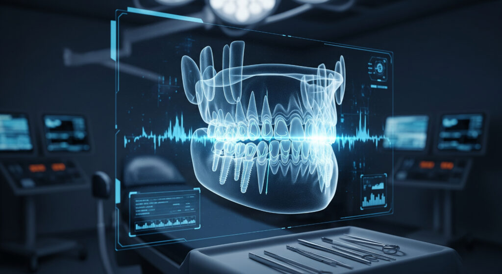
Illustration of AI-assisted analysis. Final diagnosis is always made by the dentist after a clinical examination.
AI image analysis
See how AI reads your scan before the doctor decides
Every panoramic X-ray, CBCT scan and smile photo you send is analysed by AI first. It maps bone, nerves, roots and soft tissue, flags areas that need attention and prepares measurements. Dr. Abdurrahman Öztürk then reviews every finding and makes the clinical decision.
What AI 3D dental scanning actually captures
Dental imaging is not one test but a small family of tools, each answering a different question. An intraoral scanner is a slim handheld wand that projects light onto your teeth and records their surface shape and colour. It sees enamel, existing fillings, crowns and the gum line in fine detail, but it cannot see inside a tooth or through the gums. That is the job of X-ray based imaging, which shows roots, bone and hidden decay.
CBCT, short for cone beam computed tomography, is the three-dimensional member of the X-ray family. Instead of a single flat picture, it produces a volume you can slice in any direction: front to back, side to side or top to bottom. At AIDENT Clinic the AI layer sits on top of all of these images, helping to organise, segment and highlight what they contain, while the dentist decides which images you genuinely need and what they show.
Intraoral 3D scan
A light-based scan of tooth and gum surfaces that replaces putty impressions. It involves no radiation and can be repeated whenever a fresh record is useful.
CBCT 3D X-ray
A rotating X-ray capture that rebuilds the jaw in three dimensions, used when bone volume, root shape or nerve position must be known precisely.
2D digital X-rays
Panoramic and small intraoral X-rays remain useful for screening decay, checking fillings and general overviews with a lower dose than 3D imaging.
Inside the AI intraoral scan: what to expect in the chair
Many patients remember traditional impressions as the least pleasant part of dental treatment: a tray of thick paste held in the mouth until it sets, sometimes triggering gagging and occasionally needing to be repeated. A digital intraoral scan avoids that entirely. You sit back comfortably, the teeth are gently dried, and the dentist or assistant glides the scanner wand over each surface while a 3D model builds live on the screen beside you.
Because the model appears in real time, missing areas are visible immediately and can be rescanned on the spot rather than discovered days later in a laboratory. AI-assisted software helps here by flagging holes in the surface, distinguishing teeth from cheeks and tongue, and matching upper and lower arches into your natural bite. Patients with a sensitive gag reflex usually find the process far easier, since the wand is small and you can pause whenever you need to swallow or rest.
- No impression paste and no waiting for material to set
- You can watch your own teeth appear in 3D on the screen
- Short breaks are possible at any point during the scan
- Upper arch, lower arch and bite are recorded as one set
- The file can be shared digitally with the lab the same day
AI CBCT imaging: seeing bone, roots and nerves in 3D
For a CBCT scan you stand or sit still while the machine arm rotates once around your head. The exposure itself lasts only seconds, and the whole appointment, including positioning, is short. You will be asked to remove metal items such as earrings, glasses, hairpins and removable dentures, because metal can create bright streaks across the image. A bite block and head support help you stay still, which matters because movement blurs a 3D scan.
Once reconstructed, the CBCT volume lets the dentist measure bone height and width, follow the course of the lower jaw nerve, see how close upper roots sit to the sinus floor and inspect infections at root tips that a flat X-ray can hide behind overlapping structures. AI segmentation software can outline these structures in colour, which speeds up the reading and makes the images far easier to explain to you during your consultation.
Field of view
A CBCT can cover a few teeth, one jaw or the whole face. The smallest field that answers the clinical question is chosen, which also keeps the dose lower.
What CBCT does not show well
Soft tissues such as gums, muscles and the tongue appear poorly on CBCT. That is one reason the intraoral surface scan is often fused with it.
Radiation, safety and why not everyone needs a 3D X-ray
Intraoral scanning uses light only, so it carries no radiation at all. CBCT does use X-rays. As a general rule, a dental CBCT gives a higher dose than a panoramic or small intraoral X-ray, but usually a lower dose than a conventional medical CT of the head. The exact dose depends on the machine, the settings and how large an area is scanned, which is why no single number applies to every patient.
AIDENT follows the ALARA principle, meaning radiation is kept as low as reasonably achievable. A CBCT is taken only when the answer will change your treatment, such as implant planning, impacted wisdom teeth, complex root canal anatomy or bone assessment before surgery. If you are pregnant or think you may be, tell the team before any X-ray; non-urgent imaging is normally postponed. Recent scans you already have may be reused when their quality and timing are adequate.
- Intraoral 3D scans: no ionising radiation
- CBCT only when it adds clinically useful information
- Smallest suitable field of view selected for each case
- Pregnancy, children and repeat scans assessed individually
What AI adds to dental imaging, and where its limits are
A single CBCT volume can contain hundreds of slices. Reading it thoroughly takes time and concentration, and important findings can sit in an area unrelated to the reason for the scan. AI software helps by automatically separating teeth and bone, marking the nerve canal, highlighting dark areas that may represent infection or bone loss, and lining up the scan with your intraoral model. This gives the dentist an organised starting point and a second set of eyes on every image.
AI is not a diagnosis machine, though. It can miss findings, overmark harmless variations and struggle with images blurred by movement or metal. Its output is a suggestion that must be checked. At AIDENT Clinic, Dr. Abdurrahman Öztürk reviews the raw images himself, confirms or rejects every AI marking and relates the images to your symptoms and clinical examination before any conclusion is drawn.
Sending scans before you travel and taking your data home
Many AIDENT patients live outside Turkey, so imaging is planned around travel. Before you book flights, you can send clear photos of your smile and any existing X-rays or CBCT files on WhatsApp. These give an initial impression of what new imaging might be needed. Final decisions always wait for the in-person examination and fresh scans in Istanbul, because older or lower-quality images cannot always be relied on for precise planning.
After your visit, your digital records belong to your care. CBCT volumes are typically stored in the standard DICOM format, and intraoral scans are commonly exported as STL or similar mesh files, which most modern dental practices can open. Ask for copies before you leave so that your dentist at home has the same 3D information for follow-up, maintenance or any future treatment.
- Share photos and old X-rays on WhatsApp before travelling
- New scans are usually taken early in your Istanbul stay
- Ask for your CBCT (DICOM) and scan files (STL) on departure
Conventional vs AI-assisted
Traditional dental records vs AI 3D scanning at AIDENT
Who it suits
Who benefits most from AI 3D dental imaging?
- Patients planning dental implants who need bone volume and nerve position assessed
- Anyone needing crowns, veneers or bridges who wants mould-free digital impressions
- Patients with impacted or awkwardly placed wisdom teeth before extraction
- People with persistent tooth pain where a flat X-ray did not explain the cause
- Orthodontic and aligner patients who need an accurate starting record of their bite
- International patients who want complete digital records to take home
Your treatment journey
Your AI imaging journey with AIDENT
Before travel
Online review
Send smile photos and any existing X-rays or scans on WhatsApp so the team can suggest which new images are likely to be needed.
Day 1
Examination and scans
Clinical exam, intraoral 3D scan and, only where justified, a CBCT or 2D X-ray taken at the clinic.
Day 1–2
AI processing
Images are reconstructed, segmented and fused by AI software, preparing an organised dataset for reading.
Day 2
Doctor reading and consultation
Dr. Öztürk reviews every slice and marking, then walks you through your 3D images and the options they reveal.
Departure
Records to take home
You receive copies of your scan files so your home dentist can continue care with the same information.
Why patients choose it
Comfort
No impression paste, short chair time and the freedom to pause make digital scanning far easier for most patients.
Detail in three dimensions
CBCT removes the overlap of flat X-rays, revealing roots, bone and nerve canals as they actually sit in the jaw.
Fewer repeat visits
Live scanning means incomplete records are corrected in the same appointment instead of after a lab rejects them.
Organised AI reading
Segmentation and markings give the dentist a structured overview, while he still reads every image himself.
Clear explanations
Seeing your own jaw in 3D makes it easier to understand why a treatment is or is not recommended.
Portable records
Standard digital files travel with you, supporting follow-up care wherever you live.
Your doctor
Every scan read by Dr. Abdurrahman Öztürk
Dr. Abdurrahman Öztürk holds a PhD in dentistry and oral and maxillofacial surgery and brings more than 15 years of experience in dentistry and dental surgery, having trained at a leading Istanbul university. Surgical training matters in imaging: reading a CBCT means understanding nerves, sinuses and bone the way a surgeon must. At AIDENT Clinic, AI software prepares and marks your images, but Dr. Öztürk personally reviews each intraoral scan and CBCT volume, checks the AI findings against your examination and makes every clinical decision. AI supports the reading; the doctor decides what it means.
Aftercare
After your scan: practical notes
- There is no recovery time; you can eat, drink and travel normally straight after scanning
- Ask for your CBCT and intraoral files before leaving Istanbul and keep them backed up
- Share the files with your home dentist so future X-rays can be compared with them
- Avoid unnecessary repeat CBCTs elsewhere; show your recent scan first
- Tell any future dentist the date of your last CBCT to help them judge whether a new one is needed
- Contact the clinic on WhatsApp if you have questions about what your images showed
Related AI treatments
Questions and answers
What patients ask about AI 3D Scanning & CBCT
How long does an AI 3D dental scan take?
An intraoral scan usually takes only a few minutes per arch, and a CBCT exposure lasts just seconds. Including positioning and checking image quality, most imaging appointments are short. The AI processing and the doctor's reading happen afterwards, and your results are normally explained to you during the consultation on the same or the following day.
What is the difference between a CBCT scan and a panoramic X-ray?
A panoramic X-ray is a single flat image of both jaws, while a CBCT builds a full 3D volume. The panoramic is useful for an overview with a lower dose. CBCT shows bone width, root shape and nerve position that a flat image cannot measure, so it is reserved for cases where that extra detail changes the treatment plan.
Can I have a CBCT scan if I am pregnant?
Non-urgent dental X-rays, including CBCT, are generally postponed during pregnancy. Always tell the team if you are pregnant or might be. Intraoral 3D scanning uses only light, so it can still be used for records. If an urgent dental problem needs imaging, the dentist will weigh the need carefully and use protective measures.
Does AI really make dental scans more accurate?
AI does not change what the scanner or X-ray machine physically captures, but it helps make better use of the images. It can flag gaps in an intraoral scan, reduce noise, segment teeth and nerves and highlight suspicious areas. That supports a more thorough reading, while accuracy still depends on good capture and on the dentist checking every result.
Will metal crowns or fillings ruin my CBCT scan?
Metal will not ruin a CBCT, but it can cause bright streaks and shadows near the restoration. Removable metal items are taken out before scanning, and AI-assisted artefact reduction can make images cleaner. Where streaks still hide an area, the dentist combines CBCT with your intraoral scan, 2D X-rays and the clinical examination.
Can I get my 3D dental scan files to take home?
Yes, you can ask for copies of your imaging before you leave Istanbul. CBCT volumes are usually provided in the standard DICOM format and intraoral scans as STL or similar files. Most modern dental practices can open them, which helps your home dentist continue your care with the same detailed information.
How much time do I need in Istanbul for 3D dental imaging?
For imaging alone, one appointment is usually enough, and results can be discussed the same or next day. Most international patients combine scanning with treatment, so the full stay depends on what the images reveal. Sending photos and old X-rays on WhatsApp first helps the team estimate your timetable before you book.
Is a CBCT scan safe for children?
CBCT can be used for children when there is a clear clinical reason, such as unerupted or impacted teeth, but it is not a routine test. Children are more sensitive to radiation, so the smallest suitable field of view and lowest practical settings are chosen. Often a 2D X-ray or an intraoral scan answers the question instead.
Can an intraoral scanner replace all dental impressions?
For most crowns, bridges, veneers, aligners and implant restorations, a digital scan replaces traditional impressions. Some situations, such as very deep margins under the gum or certain complete denture cases, may still need a conventional impression or a combined method. The dentist chooses the technique that gives the most reliable record for your case.
What should I do to prepare for a CBCT scan?
No special preparation is needed beyond removing metal items. Take out earrings, piercings, hairpins, glasses and removable dentures or retainers before the scan. Wear clothes without metal near the neck if possible, mention any pregnancy, and stay completely still during the few seconds the arm rotates, because movement blurs the image.
Can AI find problems in my X-rays that a dentist might miss?
AI can draw attention to areas that deserve a closer look, which is helpful in large 3D scans. It is a support tool, not a replacement for clinical judgement. AI can also mark harmless features or miss real problems, so at AIDENT every finding is checked by Dr. Öztürk against the raw images and your examination.
Can I send my existing CBCT for an online opinion before travelling?
Yes, you can share an existing CBCT or X-rays on WhatsApp, ideally with the original files rather than screenshots. They help with an early assessment of your options. Whether they are good enough for final planning depends on their quality and age, so a new scan in Istanbul may still be recommended.
Who is the best dentist for AI 3D dental scanning in Istanbul?
The best choice is a dentist who combines modern 3D imaging with the clinical experience to read it carefully. At AIDENT Clinic, Dr. Abdurrahman Öztürk, with a PhD in oral and maxillofacial surgery and over 15 years of experience, personally reviews every AI-processed scan and makes all clinical decisions.
Is the intraoral scanner light harmful to my eyes or teeth?
No, the scanner uses visible light similar to normal dental lighting and is designed for repeated clinical use. It does not heat or damage teeth, and it emits no X-rays. You may be offered protective glasses for comfort, and you can close your eyes during the scan if the light bothers you.
No question matches your search. Ask us directly on WhatsApp.
Start with an AI review of your smile
Send photos of your teeth and any X-rays or CBCT files you already have. Our team will tell you which 3D scans are likely to be useful and how imaging fits into your treatment in Istanbul, with every case reviewed by Dr. Abdurrahman Öztürk.
Get my free AI assessment on WhatsAppFree initial review of your photos and X-rays. Reply on WhatsApp.