AI-Planned Endodontic Microsurgery
AI Apicoectomy in Istanbul: Precise Surgical Root-Tip Treatment That Saves Your Tooth
When infection persists at the very tip of a root despite a good root canal, an AI apicoectomy lets us reach the problem directly through the gum, remove the diseased root end and seal it, using AI-analyzed 3D scans to plan every millimetre under the supervision of Dr. Abdurrahman Öztürk.
 Medically reviewed by Dr. Abdurrahman Öztürk, PhD, oral & maxillofacial surgery
The model shows a root-treated tooth with the apical section removed and a root-end filling placed, surrounded by the bone window through which the surgeon works. AI segmentation of the CBCT makes these structures visible before surgery.
Short answer
What is an AI apicoectomy?
An AI apicoectomy is a minor surgical procedure in which the infected tip of a tooth root is removed through a small opening in the gum and the root end is sealed with a biocompatible filling. AI software analyzes the CBCT scan to map the lesion, root shape, nerves and sinus, and the dentist plans and performs the surgery.
- Anaesthesia
- Local, tooth area only
- Chair time
- Often 30–90 minutes
- Stitches out
- Usually after 5–7 days
- Bone healing check
- X-ray follow-up over months
AI
How AI prepares your apicoectomy before the first incision
An apicoectomy happens in a space of only a few millimetres, close to nerves, the sinus and neighbouring roots. That is why most of the work happens before surgery, in the planning stage, where AI tools help Dr. Öztürk see the root tip in three dimensions.
- 1
Case intake on WhatsApp
You send existing X-rays, previous root canal records and a description of your symptoms. A first AI-supported review tells us whether surgery is likely to be considered at all.
- 2
CBCT scan in Istanbul
A cone-beam CT gives a 3D image of the tooth, the root tip and the surrounding bone. It is the key examination for planning root-end surgery.
- 3
AI segmentation of root and lesion
Software outlines the root, the canal filling, the dark area of bone loss at the apex and nearby structures, so their size and position can be measured.
- 4
Distance and access analysis
The plan measures how far the root tip lies from the mandibular nerve, the mental foramen or the maxillary sinus, and how thick the outer bone wall is over the lesion.
- 5
Surgical plan review by the doctor
Dr. Öztürk decides whether apicoectomy is the right option, where to make the flap, how much root to resect and whether a retreatment should come first.
- 6
Follow-up comparison
Later control X-rays or scans can be compared with the original images to follow how the bone defect fills in over the healing period.
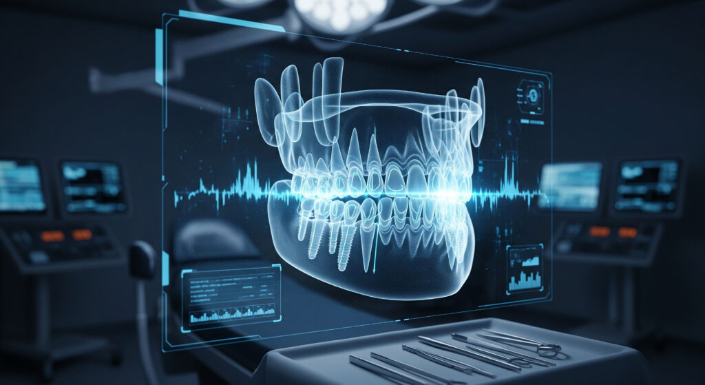
Illustration of AI-assisted analysis. Final diagnosis is always made by the dentist after a clinical examination.
AI image analysis
See how AI reads your scan before the doctor decides
Every panoramic X-ray, CBCT scan and smile photo you send is analysed by AI first. It maps bone, nerves, roots and soft tissue, flags areas that need attention and prepares measurements. Dr. Abdurrahman Öztürk then reviews every finding and makes the clinical decision.
What an apicoectomy is and when AI-planned surgery is the right step
An apicoectomy, also called root-end resection or surgical root treatment, removes the last few millimetres of a tooth root together with the inflamed tissue that surrounds it. Instead of going through the crown, as in a normal root canal, the dentist reaches the root tip from the side, through the gum and a small window in the bone. The cut root surface is then cleaned and sealed from below so that bacteria trapped in the canal can no longer leak into the jaw.
It is usually considered when a tooth has already had root canal treatment, yet a lesion at the apex will not heal, or when going back through the crown is impractical. Typical situations include a tooth with a well-fitting crown or a long post that would be damaged if removed, a root with a complex shape or a calcified section that instruments cannot pass, a persistent cyst-like lesion, or a broken file lodged near the tip. With AI apicoectomy planning we first check on the 3D scan whether surgery or another approach makes more sense for your specific tooth.
- Lesion at the root tip that persists after an adequate root canal
- Crown, bridge or post that should not be removed
- Blocked, curved or calcified canal near the apex
- Instrument fragment or filling material pushed beyond the root
- Suspected root-tip cyst that needs tissue for examination
Why the root tip is so hard to treat, and what AI shows us
The final part of a root is anatomically the most complicated. It often contains tiny side branches and accessory canals that no file can fully clean from the crown. Bacteria and their by-products can survive there and keep a chronic inflammation going, which appears on X-rays as a dark halo around the apex. This is why even a carefully performed root canal sometimes does not lead to complete healing.
A flat panoramic or periapical X-ray can hide the true size and direction of such a lesion, because it squeezes three dimensions into two. On a CBCT, AI-assisted segmentation can separate the root, the bone defect and the outer cortical plate, so the dentist sees whether the lesion has broken through the bone, whether it reaches a neighbouring root, and how the root tip is angled. These details decide where the surgical window should go and how large it needs to be.
Two-dimensional X-ray versus 3D scan
A periapical X-ray remains useful for quick checks, but for planning root-end surgery, especially on molars and premolars, a 3D scan gives information a flat image simply cannot provide, such as the buccal and palatal position of each root.
What AI does not decide
The software highlights and measures structures; it does not choose the treatment. Interpretation of the images and every surgical decision remain with Dr. Abdurrahman Öztürk after he has examined you in person.
Step by step: what happens during an AI-guided apicoectomy in Istanbul
The procedure is performed under local anaesthesia, so you stay awake but the area is numb. The dentist makes a small incision in the gum and gently lifts a flap to expose the bone over the root tip. Using the measurements taken from the AI-analyzed scan, a small opening is made in the bone exactly where the apex is expected, which helps keep the access as conservative as possible.
The inflamed tissue around the root end is removed, and in general dental practice about three millimetres of the root tip are resected, since that is where most of the hard-to-clean side canals sit. A short cavity is then prepared in the cut root end, typically with fine ultrasonic tips, and filled with a biocompatible root-end material, such as a bioceramic or MTA-type cement, that seals the canal from below. Magnification helps the surgeon inspect the cut surface for cracks or missed canals. The flap is repositioned and closed with fine stitches.
- Local anaesthesia and small gum incision
- Conservative bone window guided by 3D measurements
- Removal of the lesion and resection of the root tip
- Root-end cavity preparation and biocompatible seal
- Stitches, cold compress advice and written aftercare
Healing after AI apicoectomy: the first days and the following months
Most people describe the first two or three days as the most uncomfortable part, with tenderness, mild swelling and sometimes bruising of the cheek or lip. These signs are a normal response of soft tissue to surgery and usually settle with cold compresses, the prescribed or recommended pain relief and rest. Stitches are commonly removed after about five to seven days, at which point the gum has already closed.
The bone takes much longer. The dark area seen on the X-ray fills in gradually over many months, so success cannot be judged on the day you fly home. We therefore plan control X-rays, and where needed a follow-up scan, at intervals agreed with you. Patients abroad can have these images taken locally and send them to us; the new images can then be compared with the original AI-segmented scan to follow how the defect is filling in.
Signs that healing is on track
Pain that steadily decreases, swelling that peaks early and then fades, and no return of the gum boil or pressure you had before surgery are reassuring signs during the first weeks.
When to contact us
Swelling that keeps increasing after the third day, fever, persistent bleeding, or numbness that does not improve should be reported to the clinic on WhatsApp straight away.
Risks, limits and honest expectations of root-end surgery
An apicoectomy is a well-established procedure, but it is still surgery and it cannot guarantee that a tooth will be saved. The outcome depends on factors such as the quality of the existing root filling, whether a crack is present in the root, the amount of bone support, and the position of the tooth. A vertical root fracture discovered during surgery, for example, usually means the tooth cannot be kept, and we would then discuss extraction and replacement with you.
Specific risks depend on location. Lower premolars and molars may lie close to the nerve that supplies feeling to the lip and chin, so temporary or, rarely, longer-lasting numbness is possible. Upper back teeth can sit near the maxillary sinus, which may be opened during surgery and usually heals with simple precautions. Gum recession at the incision line, infection and the need for a further procedure are other possibilities. AI distance analysis helps us anticipate these risks, and in some cases it shows that a different treatment is the safer choice.
- Temporary swelling, bruising and tenderness
- Possible numbness near the lower nerve
- Possible sinus involvement for upper molars
- Slight gum recession around crowns at the front
- Persistent lesion that may need further treatment
Apicoectomy, retreatment or implant: how the AI review helps you decide
Root-end surgery is one of several ways to deal with a failing root canal, and it is not always the first. If the old root filling is short, poorly condensed or a canal was missed, cleaning the canals again from the crown often addresses the cause more completely, and surgery may only be needed later if healing still does not happen. If the root is cracked or there is too little tooth structure left, extraction followed by an implant or bridge may offer a more predictable result.
During the AI-supported review, the scan, the quality of the existing treatment and the restoration on top of the tooth are assessed together. Dr. Öztürk then explains which options are realistic for your tooth, what each involves and what the main uncertainties are. For many patients, keeping their own tooth with an apicoectomy is preferable because it preserves the natural root, the surrounding bone and the gum contour, but this is a decision we make together after the clinical examination.
Planning an AI apicoectomy trip to Istanbul
Because the surgery itself is usually done in one appointment, an apicoectomy fits well into a short stay in Istanbul. A typical plan includes a consultation and CBCT on arrival, surgery on the same or the next day, a check-up within a few days and stitch removal before you leave, which is why around a week is generally recommended. If a retreatment or a new crown is needed as well, the schedule becomes longer and we will discuss it in advance.
You can start from home: send your X-rays, any CBCT you already have, and a short description of your symptoms to our WhatsApp number. A preliminary AI-supported assessment helps us tell you whether surgery is likely to be needed and which records to bring. Avoid flying on the same day as surgery when possible, and plan a calm day afterwards rather than intensive sightseeing.
Conventional vs AI-assisted
Conventional apicoectomy planning versus AI-assisted planning at AIDENT
Who it suits
Who may be a good candidate for AI apicoectomy
- Patients with a root-treated tooth whose lesion at the apex has not healed
- People who have a good crown or bridge they do not want removed
- Teeth with a long post or a blocked canal that limits retreatment
- Cases with a broken instrument or excess filling beyond the root tip
- Patients with a suspected root-tip cyst that needs removal and examination
- Adults in good general health with enough bone and gum support around the tooth
Your treatment journey
A typical AI apicoectomy timeline
Before travel
Online review
You send X-rays and symptoms on WhatsApp and receive an initial opinion on whether surgery is likely.
Day 1
Examination and CBCT
Clinical examination, CBCT scan and AI-assisted analysis, followed by a planning discussion with Dr. Öztürk.
Day 1–2
Apicoectomy
Surgery under local anaesthesia, usually completed in a single appointment, with written aftercare instructions.
Day 3–4
Early check
The clinic checks swelling, comfort and the incision line and answers your questions.
Day 5–7
Stitch removal
Sutures are removed once the gum has closed, and you are cleared to travel home.
Following months
Bone healing follow-up
Control X-rays taken locally or in Istanbul are compared with the original scan.
Why patients choose it
Keeps your natural tooth
When it succeeds, an apicoectomy preserves your own root, the bone around it and your bite, instead of replacing the tooth.
Existing crown can stay
Because access is through the gum, a well-made crown, bridge or post does not have to be removed.
3D-informed access
AI segmentation of the scan helps position the bone window precisely and keep it small.
Safer around nerves and sinus
Measured distances to the nerve and sinus help the surgeon plan with those structures in mind.
Tissue can be examined
Lesion tissue removed during surgery can be sent for examination when the diagnosis is uncertain.
Compact treatment schedule
Surgery is usually a single visit, making it practical for international patients staying about a week.
Your doctor
Your apicoectomy is planned and performed under Dr. Abdurrahman Öztürk
Dr. Abdurrahman Öztürk holds a PhD in dentistry and oral and maxillofacial surgery and has more than 15 years of experience in dental surgery. He graduated from a leading Istanbul university and supervises every case at AIDENT Clinic. For apicoectomy, that surgical background matters: the AI-segmented CBCT shows him the lesion and the nearby nerve or sinus, but it is his clinical judgement that decides whether surgery is indicated, how the flap is designed and how much root is removed. AI supports the planning; Dr. Öztürk examines you, explains the options honestly and makes every decision.
Aftercare
Aftercare following your apicoectomy
- Apply a cold compress to the cheek in short intervals during the first day
- Take pain relief and any prescribed medication exactly as instructed
- Eat soft, lukewarm food and chew on the other side for several days
- Do not pull the lip to look at the stitches, and avoid touching them with your tongue
- Avoid smoking, alcohol and hard physical exercise while the gum heals
- Brush the other teeth normally and clean near the area gently as advised
- Keep your follow-up X-ray appointments, at home or in Istanbul
Related AI treatments
Questions and answers
What patients ask about AI Apicoectomy
Is an apicoectomy painful?
The surgery itself should not be painful because the area is fully numbed with local anaesthetic. You may feel pressure or vibration. Afterwards, tenderness and swelling are common for two or three days and are usually controlled with cold compresses and standard pain relief. Most patients return to light daily activities the next day.
How long does an apicoectomy take?
Many apicoectomies take between thirty and ninety minutes. Front teeth with easy access are often quicker, while premolars and molars with several roots or a position close to the nerve or sinus take longer. The AI analysis of your CBCT helps us estimate the time in advance and tell you before the appointment.
How long does it take to heal after an apicoectomy?
The gum usually closes within about a week, which is when stitches are removed. Bone healing is slower: the dark area on the X-ray gradually fills in over many months. That is why we compare follow-up X-rays with your original AI-analyzed scan rather than judging success immediately.
How many days do I need to stay in Istanbul for an apicoectomy?
Around a week is generally recommended. That allows time for the examination and CBCT, the surgery, an early check and stitch removal before you fly. If you also need a root canal retreatment or a new crown, the stay may be longer, and we will plan the dates with you beforehand.
Does AI really make an apicoectomy safer?
AI does not perform the surgery, but it can make planning more precise. Segmentation of the CBCT shows the size and direction of the lesion and measures distances to the nerve and sinus. This helps the dentist choose a conservative access and anticipate risks. Every result is reviewed and every decision is made by Dr. Öztürk.
Who is the best dentist for AI apicoectomy in Istanbul?
Look for a dentist with real surgical training, access to CBCT imaging and a clear follow-up plan. At AIDENT Clinic, apicoectomies are planned with AI-assisted 3D analysis and performed under Dr. Abdurrahman Öztürk, who holds a PhD in oral and maxillofacial surgery and has over 15 years of experience.
Is an apicoectomy better than pulling the tooth and placing an implant?
It depends on the tooth. If the root is intact and has enough bone support, an apicoectomy can keep your natural tooth, which many patients prefer. If the root is cracked or badly weakened, an implant may be more predictable. After the AI-supported scan review, Dr. Öztürk explains which option suits your situation.
Can I have an apicoectomy on a back molar?
Yes, apicoectomy is possible on many molars, but it is more demanding. Lower molars can be close to the nerve and have thick bone, and upper molars may lie near the sinus. A CBCT with AI measurements shows whether access is safe; in some cases retreatment or another option is recommended instead.
Will I have a scar or visible stitches after an apicoectomy?
There is no scar on the face because the incision is inside the mouth. Fine stitches in the gum are visible only when you lift your lip and are removed after about a week. Around front crowns, a small change in the gum line can occasionally occur, and we design the flap to reduce this risk.
Can an apicoectomy be done more than once on the same tooth?
A repeat apicoectomy is sometimes possible, but each surgery shortens the root a little further, so the tooth must still have enough length and bone support. A new CBCT and AI analysis help us see why the first surgery did not heal and whether repeating it, retreating the canal or replacing the tooth is the more sensible choice.
What should I eat after an apicoectomy?
Choose soft, lukewarm foods such as yoghurt, soups that are not hot, eggs, mashed vegetables and pasta for the first few days. Chew on the opposite side and avoid crunchy, spicy or very hot foods, as well as using a straw. You can gradually return to a normal diet as tenderness fades.
Can I fly home right after an apicoectomy?
Flying on the same day is best avoided when possible, mainly so that bleeding and swelling can be checked first. Most patients travel after their stitches are removed. Upper molar cases in which the sinus was involved may need specific advice about pressure changes, which Dr. Öztürk will give you personally.
Can I send my X-rays on WhatsApp to find out if I need an apicoectomy?
Yes. Send your existing X-rays, any CBCT and a short description of your symptoms to +90 534 445 9444. We will give you a preliminary AI-supported opinion on whether surgery is likely. A final decision is only possible after a clinical examination and a new 3D scan in Istanbul.
No question matches your search. Ask us directly on WhatsApp.
Find out if an AI apicoectomy can save your tooth
Send your X-rays and a description of your symptoms on WhatsApp. We will review them with AI-supported analysis, and Dr. Abdurrahman Öztürk will tell you honestly whether root-end surgery, retreatment or another option is the best way forward.
Get my free AI assessment on WhatsAppFree initial review of your photos and X-rays. Reply on WhatsApp.